Treatment of adult patients with wet (wet) age-related macular degeneration (AMD) with predominantly classical subretinal choroidal neovascularization (CNV) or adult patients with subfoveal choroidal neovascularization in the course of high myopia.
Composition:
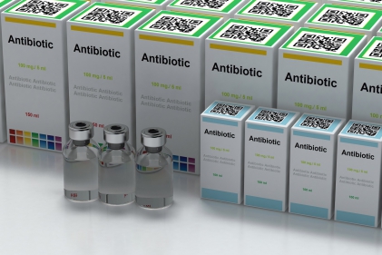
One vial contains 15 mg of verteporfin.
Action:
Verteporfin, also referred to as benzoporphyrine derivatives (BPD-MA), consists of a mixture of equally active BPD-MA isomersC and BPD-MAD in a 1: 1 ratio. It is used as a light-activated drug (sensitizing to light). Activation generates an unstable, highly reactive oxygen in the atomic form, causing damage to biological structures in the area to which it can diffuse, leading to local vascular occlusion, cell damage, and under certain conditions to cell death. The selectivity of photodynamic therapy (PDT) with verteporfin is based on localized exposure to light, and on selective and rapid uptake and retention of verteporfin by rapidly dividing cells, including endothelial cells of neovascular choroidal vessels. The maximum concentration of the drug in the blood after a 10-minute infusion, at a dose of 6 mg / m2 pc. or 12 mg / m2 pc, was 1.5 μg / ml and 3.5 μg / ml, respectively. 90% of verteporfin is bound to plasma proteins and 10% to blood cells, a very small portion of which is associated with cell membranes. The verteporfin ester group is hydrolyzed by plasma and hepatic esterases, resulting in the formation of a benzoporphyrin derivative (BPD-DA). BPD-DA is also a photosensitizer, but its systemic exposure is small. The half-life of verteporfin in the elimination phase from plasma is 5-6 h. The drug is excreted in bile.
Contraindications:
Hypersensitivity to verteporfin or other components of the preparation. Porphyria. Severe liver dysfunction.
Precautions:
Exercise caution in patients with moderate hepatic impairment or biliary obstruction - lack of experience in the treatment of such patients; increased exposure to verteporfin is possible. Do not use the product again in patients who have a significant decrease in vision (equivalent to 4 or more lines) within one week of the first course of treatment, at least until the vision improves before treatment; the potential benefits and risks of further treatment should be carefully considered. If extravasation occurs, the infusion should be stopped immediately. In order to avoid extravasation, before entering the monitored infusion, attach the intravenous access to the largest available forearm vein to allow free flow, preferably to the median elbow flexion. It is not recommended to insert the cannula into small veins on the back of the hand. Patients should be under strict medical supervision during the infusion. Caution should be exercised when considering the use of the preparation in patients under general anesthesia - lack of experience in the treatment of such patients. There are no clinical data indicating the advisability of simultaneous treatment of the other eye. However, if the treatment of the other eye is necessary, it should be irradiated immediately after irradiation of the first eye, but no later than 20 minutes after the start of the infusion. The preparation is not indicated in children and adolescents - the safety and efficacy in this group of patients has not been established. The preparation contains butylhydroxytoluene, which can be irritating to the eyes, skin and mucous membranes.
Pregnancy and lactation:
It must not be used during pregnancy unless clearly necessary. Do not give the preparation to nursing mothers or stop breast-feeding for 48 hours after its administration.
Side effects:
Common: hypercholesterolemia, reduced visual acuity (sometimes significant), blurred vision or flashes, visual field defects (scotoma, gray or dark capsule and black spots), nausea, photosensitivity reaction, injection site reactions (pain, edema) , inflammation, haemorrhage), weakness,reactions related to intravenous infusion, mainly presented as back pain. Uncommon: hyperesthesia, retinal detachment (without tearing), subretinal / intra retinal hemorrhage, intravitreal haemorrhage, hypertension, hypersensitivity at the injection site, injection site haemorrhage, discoloration at the injection site, fever, pain. Rare: no perfusion in the retinal or choroidal vessels. Not known: vasovagal reactions and hypersensitivity reactions that may rarely be serious (general symptoms may include headache, malaise, fainting, hyperhydration, dizziness, rash, urticaria, pruritus, shortness of breath, redness, changes in blood pressure and heart rate), retinal pigment epithelial detachment, macular edema, retinal edema, myocardial infarction (mainly in patients with a history of cardiovascular disorders, sometimes within 48 hours after infusion), bladder at the injection site, infusion associated with pain in the chest. Chest pains and back pain caused by the way the medicine is given (infusion) may radiate to other parts of the body (including the pelvis, shoulder and ribs).
Dosage:
Adults (including the elderly). Photodynamic therapy takes place in two stages.The first step: 10-minute intravenous infusion of the preparation at a dose of 6 mg / m2 pc, diluted in 30 ml of solution for infusion. The preparation should be dissolved in 7 ml of water for injections to obtain 7.5 ml of a 2 mg / ml solution. In order to obtain a dose of 6 mg / m2 Dilute the appropriate amount of the solution with 5% Glucose for injection to a final volume of 30 ml. Do not use NaCl solution. After reconstitution and dilution, the solution should be protected from light until administration; the diluted solution should be administered within a maximum of 4 hours after preparation.The second stage: activation of the preparation with light exactly 15 minutes after the start of the infusion. For this purpose, a diode laser is used, which produces red light without thermal influence (wavelength 689 nm +/- 3 nm). At a recommended light intensity of 600 mW / cm2, the required light dose 50 J / cm2 is delivered within 83 s. The largest linear dimension of the subretinal neovascularization lesion is determined by fluorescein angiography and fundus photography. It is recommended to use the cameras for examination of the fundus with magnification in the range of 2.4-2.6 times. The focus of treatment should include all neovascular changes, blood and / or blocked fluorescence. To provide treatment for poorly marked change boundaries, add 500 μm margin around the visible change. In the nasal direction, the focus of treatment must be at least 200 μm away from the temporal edge of the optic disc. The maximum focal size used in the first treatment cycle in clinical trials was 6400 μm. In the treatment of lesions greater than the maximum size of the focus of treatment, light should be applied to the largest possible area of active change. In the case of relapses of blood cells penetrating the newly formed choroidal vessels, the treatment can be repeated up to 4 times a year.